
Coronary Angiography: A Comprehensive Diagnostic Procedure for Heart Health
Coronary angiography is a crucial diagnostic procedure used to evaluate the condition of the coronary arteries and detect blockages that may lead to heart disease. This minimally invasive technique provides detailed images of the blood vessels, allowing cardiologists to assess blood flow to the heart and determine the best course of treatment for patients with suspected or existing coronary artery disease (CAD).
What is Coronary Angiography?
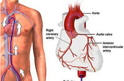
Coronary angiography is a specialized X-ray test that utilizes contrast dye and fluoroscopy to visualize the coronary arteries. The procedure helps in identifying narrowed or blocked arteries caused by plaque buildup, which can result in conditions such as angina, heart attacks, and other cardiovascular complications.
Why is Coronary Angiography Performed?
Your cardiologist may recommend coronary angiography if you experience symptoms of heart disease, including:
- Chest pain (angina)
- Shortness of breath
- Unexplained fatigue
- Abnormal results from stress tests or electrocardiograms (ECG)e
- Dizziness or fainting spells
The procedure is essential for diagnosing coronary artery disease, evaluating heart function before major surgeries, and determining the need for interventions such as angioplasty or bypass surgery.
The Procedure: What to Expect
Under the expert guidance of Dr. Madhur Jain, Senior Consultant in Cardiology at Paras Hospital, Gurugram, patients undergo coronary angiography with utmost care and precision. The process involves the following steps:
Preparation
The patient may be asked to fast for several hours before the procedure. Blood tests and ECG may be conducted to assess overall health.
Anesthesia and Catheter Insertion
Local anesthesia is administered to numb the area, usually in the wrist (radial artery) or groin (femoral artery). A thin catheter is then carefully inserted into the artery and guided toward the coronary arteries.
Injection of Contrast Dye
A special contrast dye is injected through the catheter, making the arteries visible on X-ray imaging.
Fluoroscopy Imaging
Real-time X-ray images are captured to detect any blockages, narrowing, or abnormalities in the arteries.
Completion and Recovery
Once the imaging is complete, the catheter is removed, and a bandage is applied to the insertion site. Patients are monitored for a few hours before being discharged.